|
 |
■ Bioresources information is available at the following URLs
|
 |
|
Introduction to Resource Center <No. 48> Future Perspectives and Recent Research Achievements of the National BioResource Project "Japanese monkeys"
Tadashi Isa (Professor, National Institute for Physiological Sciences,
National Institutes of Natural Sciences)
 The Japanese monkey (Macaca fuscata) is a medium-sized monkey species of the genus Macaca. There is an international trend toward not using anthropoid apes such as chimpanzees and gorillas for invasive experiments and research in the future, and macaques are the closest relatives to humans among animals that are used for experiments and research. In particular, many brain science researchers have used Japanese monkeys for their research in Japan. One reason is that the Japanese monkeys that have been caught to prevent crop raiding were provided to researchers. Other reasons include their gentleness, which makes them easy to handle as experimental animals, and their high intelligence quotient, which many researchers think makes them suitable for higher brain function research. At present, animals caught from their natural habitats are almost impossible to use for experiments and research. Because the National BioResource Project (NBRP) has established a system to supply macaques bred for research, brain science research can now be smoothly pursued. The Japanese monkey (Macaca fuscata) is a medium-sized monkey species of the genus Macaca. There is an international trend toward not using anthropoid apes such as chimpanzees and gorillas for invasive experiments and research in the future, and macaques are the closest relatives to humans among animals that are used for experiments and research. In particular, many brain science researchers have used Japanese monkeys for their research in Japan. One reason is that the Japanese monkeys that have been caught to prevent crop raiding were provided to researchers. Other reasons include their gentleness, which makes them easy to handle as experimental animals, and their high intelligence quotient, which many researchers think makes them suitable for higher brain function research. At present, animals caught from their natural habitats are almost impossible to use for experiments and research. Because the National BioResource Project (NBRP) has established a system to supply macaques bred for research, brain science research can now be smoothly pursued.
 At present, Japanese monkeys are known to have the most northern habitat of all species in the genus Macaca. Research on fossils discovered in the Japanese Islands suggests that when the Japanese Islands were connected to the Asian mainland 430,000−630,000 years ago, Japanese monkeys migrated from the region of China to the present Japanese Islands through the Korean Peninsula. Namely, macaques existed in the Korean Peninsula at that time. These macaques in the Korean Peninsula are considered to be a species related to the modern Macaca mulatta. Research on genetic backgrounds of various macaques revealed that the genetic uniformity of Japanese monkeys was higher than that of other macaques widely distributed in Southeast Asia, such as Macaca mulatta and Macaca fascicularis. Recently, it was revealed that although Macaca mulatta and Macaca fascicularis were resistant to malaria, Japanese monkeys developed fulminating malaria. This interesting finding has attracted the attention of some malaria researchers. At present, Japanese monkeys are known to have the most northern habitat of all species in the genus Macaca. Research on fossils discovered in the Japanese Islands suggests that when the Japanese Islands were connected to the Asian mainland 430,000−630,000 years ago, Japanese monkeys migrated from the region of China to the present Japanese Islands through the Korean Peninsula. Namely, macaques existed in the Korean Peninsula at that time. These macaques in the Korean Peninsula are considered to be a species related to the modern Macaca mulatta. Research on genetic backgrounds of various macaques revealed that the genetic uniformity of Japanese monkeys was higher than that of other macaques widely distributed in Southeast Asia, such as Macaca mulatta and Macaca fascicularis. Recently, it was revealed that although Macaca mulatta and Macaca fascicularis were resistant to malaria, Japanese monkeys developed fulminating malaria. This interesting finding has attracted the attention of some malaria researchers.
 In the fiscal year 2013, the NBRP expanded the distribution of its resources, namely, Japanese monkeys, from researchers in neuroscience to those in biological sciences. In response to concerns from users (researchers) that "the distribution of resources once a year is inconvenient for me," the NBRP is now open for requests twice a year and distributes them three times a year. Thus, the NBRP aims to establish a user-friendly system for its activities. Although we still cannot estimate how much the demand for resources will increase, researches who wish to compare Japanese monkeys with other macaques of the genus Macaca and those who want to use Japanese monkeys for any experiment should contact the Section of Primate Animal Models, National Institute for Physiological Sciences (former Section of NBR Promotion, 0564-55-7867, nbr-nips@nips.ac.jp). In the fiscal year 2013, the NBRP expanded the distribution of its resources, namely, Japanese monkeys, from researchers in neuroscience to those in biological sciences. In response to concerns from users (researchers) that "the distribution of resources once a year is inconvenient for me," the NBRP is now open for requests twice a year and distributes them three times a year. Thus, the NBRP aims to establish a user-friendly system for its activities. Although we still cannot estimate how much the demand for resources will increase, researches who wish to compare Japanese monkeys with other macaques of the genus Macaca and those who want to use Japanese monkeys for any experiment should contact the Section of Primate Animal Models, National Institute for Physiological Sciences (former Section of NBR Promotion, 0564-55-7867, nbr-nips@nips.ac.jp).
|
 From recent research: From recent research:
A pathway for selective and reversible neurotransmission-blocking method by using double infection of viral vectors was established using a macaque and behavior control was achieved (Kinoshita et al. (2012): Genetic dissection of the circuit for hand dexterity in primates. Nature 487: 235-238).
Humans and other higher primates are said to have explosively evolved after acquiring the ability to move fingers skillfully. The ability to skillfully move each finger individually is considered to have been acquired because the motor areas of the cerebral cortex are directly connected with the motor nerve cells in the spinal cord, which control muscles. However, in animals whose finger development is primitive and finger movement is unskillful, such as cats and rats, motion command output from the cerebral cortex is indirectly connected with the motor nerve cells in the spinal cord through interneurons in the spinal cord. This "indirect pathway" still remains in primates, but its purpose has not been clearly elucidated. Is the "old pathway" that has been left behind in the evolutionary process used in the brain of higher animals or restrained because it is obstructive? This issue has been discussed many times but has not been resolved.
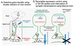
Recently, a joint research team comprising Professor Tadashi Isa and Specially Appointed Assistant Professor Masaharu Kinoshita of the National Institute for Physiological Sciences, National Institutes of Natural Sciences, Professor Kazuto Kobayashi of Fukushima Medical University, and Professor Dai Watanabe of Kyoto University combined two types of new viral vectors to develop a new method of pathway-selective gene transfer (the double-gene transfer method). By using this method, the team succeeded in selectively restraining spinal interneuronal systems (propriospinal neurons) that relay the indirect pathway (Fig. 1).
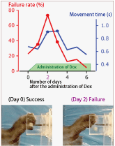
When the spinal interneuronal systems were selectively restrained, skillful movements of fingers were clearly impaired by the second day after the restraint (Fig. 2). However, functional compensation rapidly occurred through other pathways, and skillful movements of fingers had almost entirely recovered at the fifth day after the restraint. Therefore, the "indirect pathway" was revealed to play an important role in skillful finger movements. Thus, this long-term question was finally settled.
|

Fig. 1: Interruption of propriospinal neuron-selective synaptic transmissions
A) By injecting an antidromic vector (HiRet-TRE-eTeNT-EGFP) into a place where propriospinal neurons are projected and by injecting an orthodromic vector (AAV2-CMV-rtTAV16) into the intermediate zone of the central cervical spinal cord where cell bodies exist, propriospinal neurons were selectively and doubly infected with viruses
B) The double infection by itself does not cause anything to happen (left). However, when a monkey takes doxycycline (Dox), tetanus toxins are expressed by the Tet system to interrupt synaptic transmission (right). |

Fig. 2: Impairment of skillful movements of fingers when taking doxycycline (Dox)
(Graph) On the second day after the administration of Dox, the movement time was prolonged and the failure rate increased, and this continued for two days.
(Photo) Before the administration of Dox, feed could be pinched with a precision grip in which the forefinger faced the thumb. At the second day after the administration of Dox, the forefinger could not sufficiently face the thumb and the precision grip could not be pursued. |
The key element in the above research was the combination of two types of new viral vectors to develop a new technology to selectively and reversibly interrupt a specific neural circuit. Previously, a specific neural circuit could be selectively manipulated in mice, in which genetic modification was possible in the germ cells, but could not be selectively manipulated in primates. The newly developed method enabled the selective manipulation of a neural circuit in animal species, whose genetic modification had been difficult, so the method has attracted the interest of researchers worldwide. In addition, because of the above-mentioned research achievements, Professor Isa, Professor Watanabe, and Professor Kobayashi received the Commendation for Science and Technology by the Minister of Education, Culture, Sports, Science and Technology 2013.
|
|
|
Let's visualize data (using D3.js) |

Research data has become massive and complex in recent years, and data visualization techniques that use graphs and charts are becoming increasingly important to represent data that continuously changes over time, and to facilitate the quick identification of target data. In this edition, I would like to introduce "D3.js," a useful visualization tool for such scenarios.
What is D3.js ?
D3.js or Data Driven Document (http://d3js.org) is a JavaScript library for manipulating HTML documents based on data, and it facilitates the interactive representation of data. In addition, the official website (Fig. 1) has samples of complex graphs, figures, and animations, most of which can be rendered at sufficient speeds even when they are viewed on a smartphone.
| 
Fig. 1. Official website |
Let's give it a try
On the gallery website (Fig. 2), you can experiment with a variety of samples. When writing your own programs, you can refer to the source codes which are available for most examples.
Gallery website: https://github.com/mbostock/d3/wiki/Gallery
| 
Fig. 2. Gallery website |
| |
 |
Example 1:
Co-occurrence Graph
A force-directed layout is applied to co-occurrence data. This could be used, for example, for visualizing molecular networks. |
|
 |
Example 2:
Partition Graph
A partition layout can be applied to data that is stored as a tree. Visualizing categorized data in this way can have many benefits.
|
|
|

How to Use D3
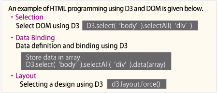
You need to include the D3.js library for manipulating HTML documents using DOM(※). In addition, you also need to define data and select a design for visualization when using D3.

※ DOM stands for Document Object Model and it is a convention for programmatically interacting with HTML elements (tags) such as <div> and <img> .


You can use DOM and D3.js to create websites which displays data that changes continuously over time. It can be difficult to use this tool initially because it involves some programming. However, once you have familiarized yourself with the tool, it is easy to create beautiful graphs and other visual representations.
(Manabu Hattori) |
|
|
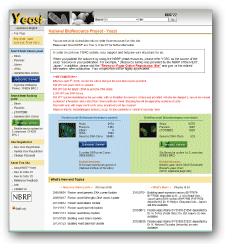
National BioResource Project " Yeast "
|

Fission yeast
・Number of strains: 9,174
・Number of genes: 62,516
Budding yeast
・Number of strains: 10,811
・Number of genes: 10,021
・Number of published papers: 615
(as of July 2013) |
|
| DB name: |
NBRP Yeast |
| URL : |
http://yeast.lab.nig.ac.jp/nig/index_en.html |
| Language: |
apanese, English |
| Original contents: |
| |
Yeast resources for research |
| ・ |
Fission yeast (strains, plasmids, libraries, full length cDNA, genomic DNA clones) |
| ・ |
Budding yeast (strains, plasmids, genomic DNA clones, gTOW6000) |
Yeast information from literature
Paper information on resource development and research achievements using resources |
| Other contents: |
| |
GenomeViewer (fission yeast, budding yeast), BLAST |
| Features: |
Resources can be ordered from the website.
Resources can be accessed from Viewer. |
| Cooperative DB: PomBase, SGD |
| DB construction group: NBRP Yeast, NBRP Information |
| Management organization: Genetic Resource Center, NIG |
| Year of DB publication: 2004 Year of last DB update: 2013 |
|
Comment from a practicing developer: The NBRP Yeast includes globally top-class information on the abundant resources conserved in the Yeast Genetic Resource Center. The number of resource species has steadily increased. We have received many resource orders and inquiries, so we feel that the NBRP Yeast is an effective database. The database working group has a plan to renew the website based on the large amount of insights accumulated during the operation of the database for nearly 10 years.
|
|
|