BioResource Newsletter Vol.5 No.1
|
|
2009/1/31
| Information on Resource-related Events |
 RIKEN BRC International Symposium, "Bacteria as Research Resources" RIKEN BRC International Symposium, "Bacteria as Research Resources"
Date: February 10 (Tue) 2009, 9:30-17:40
Place: Hyatt Regency Tokyo (2-7-2 Nishi-Shinjuku, Shinjuku-Ku, Tokyo)
 Technical training on the use of frontier bioresources Technical training on the use of frontier bioresources
"Technical Training on Culturing and Preserving Anaerobic Bacteria"
Training Period: Monday, March 2, to Tuesday, March 3, 2009 (for 2 days)
Place: Japan Collection of Microorganisms, RIKEN Wako Institute
 The 3rd International Biocuration Conference (IBC 2009) The 3rd International Biocuration Conference (IBC 2009)
Date: April 16-19, 2009, in Berlin, Germany
(for details, please visit http://projects.eml.org/Meeting2009)
Details are available at: http://www.nbrp.jp/ |
|
| Introduction to Resource Center No. 30 |
|

|
 Amphibian Resources Amphibian Resources
Yoshio YAOITA, Director and Professor, &
Masayuki SUMIDA, Professor
Institute for Amphibian Biology,
Graduate School of Science, Hiroshima University
|

|
|
|
|
|
|
 We have been involved in the development of amphibians as laboratory animals for 40 years. Since frogs living on land after metamorphosis require live feed, we have been improving the breeding administration system and developing a stable feeding system while simultaneously researching amphibians and developing strains of domestic species. Currently, we preserve and maintain approximately 30,000 wild-type and mutant amphibians of 170 strains and 50 species. In addition, we also cryopreserve 12,600 frogs of 320 groups, 112 species, 27 genera, and 9 families, which have been collected during fieldwork inside and outside Japan for approximately 30 years since 1976; we have also experimentally developed 4,000 frogs belonging to 100 special strains. We have been collecting, developing, and distributing Xenopus tropicalis under the National BioResource Project (NBRP) in collaboration with Professor Asashima’s Laboratory at the University of Tokyo since 2002. Here, we introduce some of the amphibian resources available at our institute. We have been involved in the development of amphibians as laboratory animals for 40 years. Since frogs living on land after metamorphosis require live feed, we have been improving the breeding administration system and developing a stable feeding system while simultaneously researching amphibians and developing strains of domestic species. Currently, we preserve and maintain approximately 30,000 wild-type and mutant amphibians of 170 strains and 50 species. In addition, we also cryopreserve 12,600 frogs of 320 groups, 112 species, 27 genera, and 9 families, which have been collected during fieldwork inside and outside Japan for approximately 30 years since 1976; we have also experimentally developed 4,000 frogs belonging to 100 special strains. We have been collecting, developing, and distributing Xenopus tropicalis under the National BioResource Project (NBRP) in collaboration with Professor Asashima’s Laboratory at the University of Tokyo since 2002. Here, we introduce some of the amphibian resources available at our institute.
|
|
Xenopus tropicalis
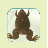
 As can be seen in Fig. 1, X. tropicalis appears to be a smaller version of Xenopus laevis, which has been frequently used for research in the field of molecular biology and developmental biology. Most of the information and knowledge accumulated about X. laevis hold true in the case of X. tropicalis, and analogous experimental techniques can also be applied to the latter. The characteristics of X. tropicalis include the following: (1) X. laevis has a tetraploid genome whereas X. tropicalis has a diploid genome, which is thus smaller in size and more suited for genetic analysis; (2) X. tropicalis is the only amphibian whose genome structure has been determined; and (3) X. tropicalis becomes sexually mature in approximately half the time that X. laevis does. X. tropicalis was inbred for more than 10 generations, because of which dermal implants are not rejected. X. tropicalis is primarily used to study early embryogenesis, organ generation, metamorphosis, and the effects of endocrine-disrupting chemicals (environmental hormones). The European Xenopus Resource Centre, which will distribute recombinant strains of X. laevis and X. tropicalis, has been established in the UK. Plans to establish a similar institute in the USA are underway. As can be seen in Fig. 1, X. tropicalis appears to be a smaller version of Xenopus laevis, which has been frequently used for research in the field of molecular biology and developmental biology. Most of the information and knowledge accumulated about X. laevis hold true in the case of X. tropicalis, and analogous experimental techniques can also be applied to the latter. The characteristics of X. tropicalis include the following: (1) X. laevis has a tetraploid genome whereas X. tropicalis has a diploid genome, which is thus smaller in size and more suited for genetic analysis; (2) X. tropicalis is the only amphibian whose genome structure has been determined; and (3) X. tropicalis becomes sexually mature in approximately half the time that X. laevis does. X. tropicalis was inbred for more than 10 generations, because of which dermal implants are not rejected. X. tropicalis is primarily used to study early embryogenesis, organ generation, metamorphosis, and the effects of endocrine-disrupting chemicals (environmental hormones). The European Xenopus Resource Centre, which will distribute recombinant strains of X. laevis and X. tropicalis, has been established in the UK. Plans to establish a similar institute in the USA are underway.
|

Fig. 1: X. tropicalis
|
"Skelepyon"--A Transparent Frog
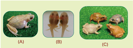
 Since the frog skin is covered with pigment cells, observing visceral organs covered by the skin is difficult. Thus, experimental animals whose organs can be observed without dissection have been needed. Last year, we successfully developed transparent frogs whose visceral organs can be observed through their skin while they are alive (Fig. 2 (A) and (B)). There are two color mutant strains of Japanese brown frogs--"black eyes," which lack iridocytes, and "gray eyes," which lack melanocytes (Fig. 2 (C))--which were used for the development of "Skelepyon." Since the frog skin is covered with pigment cells, observing visceral organs covered by the skin is difficult. Thus, experimental animals whose organs can be observed without dissection have been needed. Last year, we successfully developed transparent frogs whose visceral organs can be observed through their skin while they are alive (Fig. 2 (A) and (B)). There are two color mutant strains of Japanese brown frogs--"black eyes," which lack iridocytes, and "gray eyes," which lack melanocytes (Fig. 2 (C))--which were used for the development of "Skelepyon."
* "Skelepyon": "Skele" = Skeleton, "pyon" = hopping sound in Japanese

|
|
Fig. 2: Skelepyon
(A) An adult Skelepyon whose vessels and eggs contained in the abdomen can be observed through its transparent skin.
(B) A tadpole of Skelepyon developing legs. The development of visceral organs throughout its life can also be observed.
(C) From the top left in the clockwise direction: a wild-type Japanese red frog, a black eye, a gray eye, and a Skelepyon. |
|
 Skelepyon was developed by artificially breeding these two strains of frogs lacking iridocytes and melanocytes; as a result, Skelepyon appears transparent. Although transparent fish exist, transparent quadrupedal animals are extremely rare in nature and even in the laboratory. The visceral organs of Skelepyon can be observed from the early embryogenesis to the senescence stage of a particular individual while the individual is alive without dissection; therefore, the changes in the visceral organs during growth, the emergence and development of diseases such as cancer, and the effects of chemical substances and physical stimulations on the visceral organs and bones can easily be examined inexpensively over time. In addition, by incorporating a vector that harbors a gene encoding a fluorescent protein on the promoter region of a certain gene into the Skelepyon, "fluorescent skelepyon," which emits fluorescence upon the activation of the gene, can be developed. This technique enables the observation of real-time gene expression without requiring dissection and can thus be an extremely effective tool for gene expression analysis. It is expected that Skelepyon will become a valuable and novel experimental animal for research in environmental science, medicine, and biology. Skelepyon was developed by artificially breeding these two strains of frogs lacking iridocytes and melanocytes; as a result, Skelepyon appears transparent. Although transparent fish exist, transparent quadrupedal animals are extremely rare in nature and even in the laboratory. The visceral organs of Skelepyon can be observed from the early embryogenesis to the senescence stage of a particular individual while the individual is alive without dissection; therefore, the changes in the visceral organs during growth, the emergence and development of diseases such as cancer, and the effects of chemical substances and physical stimulations on the visceral organs and bones can easily be examined inexpensively over time. In addition, by incorporating a vector that harbors a gene encoding a fluorescent protein on the promoter region of a certain gene into the Skelepyon, "fluorescent skelepyon," which emits fluorescence upon the activation of the gene, can be developed. This technique enables the observation of real-time gene expression without requiring dissection and can thus be an extremely effective tool for gene expression analysis. It is expected that Skelepyon will become a valuable and novel experimental animal for research in environmental science, medicine, and biology.
|
|
Efficient Preservation of Endangered Species Strains and Utilization of These Resources
(Preservation and Domestication of Endangered Species for Laboratory Experiments) |
|
 Ishikawa's frogs (Fig 3 (A)) are indigenous to Amami Oshima and the capital island of Okinawa, are designated as a protected species in Kagoshima and Okinawa prefectures, and are categorized as endangered (IB) species in the Red List issued by the Ministry of the Environment. Ishikawa’s frogs exhibit pigmentation, which appears similar to golden droplets sprinkled on a green background; thus, Ishikawa’s frog is considered the most beautiful frog in Japan. The number of wild-type Ishikawa’s frogs has dramatically reduced due to environmental destruction and the overhunting of the frogs, which are domesticated as pets once captured. We obtained permission from Kagoshima and Okinawa prefectures for collecting Ishikawa’s frogs and initiated their artificial breeding in 2004 using eight pairs of male and female wild-type Ishikawa's frogs in order to preserve them in our laboratory. So far, of the artificially bred frogs, 2,000 1-4 year-old frogs have survived (Fig. 3 (B)), and the second generation has been produced during the previous breeding season. The frogs reproduced in the laboratory will be used or released for effective preservation when the number of individuals becomes so low that the frogs cannot continue existing by themselves or when the living environment deteriorates in the future. Moreover, although wild-type Ishikawa's frogs have not been used as experimental animals, the ones obtained by artificial breeding can be used for experiments effectively. Currently, we conduct several research projects using these frogs as experimental resources. Some of these studies involve the examination of the skin of Ishikawa's frogs and the identification of novel antimicrobial substances by using biochemical techniques. It is expected that the discovery of these antimicrobial substances will lead to the development of novel antibiotics.■ Ishikawa's frogs (Fig 3 (A)) are indigenous to Amami Oshima and the capital island of Okinawa, are designated as a protected species in Kagoshima and Okinawa prefectures, and are categorized as endangered (IB) species in the Red List issued by the Ministry of the Environment. Ishikawa’s frogs exhibit pigmentation, which appears similar to golden droplets sprinkled on a green background; thus, Ishikawa’s frog is considered the most beautiful frog in Japan. The number of wild-type Ishikawa’s frogs has dramatically reduced due to environmental destruction and the overhunting of the frogs, which are domesticated as pets once captured. We obtained permission from Kagoshima and Okinawa prefectures for collecting Ishikawa’s frogs and initiated their artificial breeding in 2004 using eight pairs of male and female wild-type Ishikawa's frogs in order to preserve them in our laboratory. So far, of the artificially bred frogs, 2,000 1-4 year-old frogs have survived (Fig. 3 (B)), and the second generation has been produced during the previous breeding season. The frogs reproduced in the laboratory will be used or released for effective preservation when the number of individuals becomes so low that the frogs cannot continue existing by themselves or when the living environment deteriorates in the future. Moreover, although wild-type Ishikawa's frogs have not been used as experimental animals, the ones obtained by artificial breeding can be used for experiments effectively. Currently, we conduct several research projects using these frogs as experimental resources. Some of these studies involve the examination of the skin of Ishikawa's frogs and the identification of novel antimicrobial substances by using biochemical techniques. It is expected that the discovery of these antimicrobial substances will lead to the development of novel antibiotics.■
|
|

Fig. 3: Ishikawa's Frogs
(A) An endangered Ishikawa's frog, which is considered the most beautiful in Japan (indigenous to Amami Oshima; exhibiting unique golden pigments on a green background)
(B) A two-year-old Ishikawa's frog reproduced in the laboratory
|
|
|
|

|
Erudite Lecture Series by Dr. Benno: No. 1 |
|
| |
Is your colon a flower garden ?
We have different enteric environments depending on our surroundings and our individual lifestyles. Recent studies have indicated that enteric bacteria might be responsible for various diseases such as lifestyle diseases, allergies, and psychiatric diseases. The development of a favorable enteric environment is the first step toward improved health and well-being. Let us explore the exciting world of enteric bacteria !
Surprising Hidden Strength of Enteric Bacteria
In the early 20th Century, it was believed that there are only certain types of bacteria such as Escherichia coli and Enterococci living in our gut. However, recently, various bacterial species have been identified in feces by means of molecular biological techniques, i.e., DNA analysis. It is now clear that 500?1000 bacterial species reside in the human colon and that approximately 1 trillion bacterial cells exist in 1 g of dry-weight feces. Surprisingly, the total weight of bacteria residing in the human colon is approximately 1.0?1.5 kg per person.
Enteric bacteria are deeply associated with our health. The elucidation of the functions of enteric bacteria by examining bacteria, hazardous substances, and bacterial toxins contained in feces is useful for disease suppression and controlling aging.
Association with Colon Cancer, Allergy, and Obesity!?
Colon cancer is one of the diseases caused by the presence of certain types of enteric bacteria. Although the mechanism underlying the development of colon cancer due to the presence of certain enteric bacteria has not been fully elucidated, it is believed that a sustained high-fat and high-protein diet changes the enteric environment and increases "bad" bacteria, which in turn increases the risk of colon cancer by the production of carcinogens and malignant alteration of cells. In addition, it is reported that enteric bacteria are deeply associated with allergies and obesity; thus, lately, enteric bacteria have attracted significant attention in the field of modern medicine.
Dr. Benno: Yoshimi BENNO, Head of Microbe Division,
Japan Collection of Microorganisms, RIKEN BioResource Center
|
|
|
|
 "Xen," A Server Virtualization Software
Program: Installation and Settings "Xen," A Server Virtualization Software
Program: Installation and Settings
1) Installation and Settings of Xen
An overview of "Xen," server virtualization software, was provided in the previous issue. In this issue, we discuss the installation and settings of "Xen" on CentOS 5.2 (an open-source operating system compatible with Red Hat Enterprise Linux).
2) Installation of CentOS (the host OS for Xen)
Firstly, CentOS, which is the host OS for Xen, should be installed. The image file of CentOS is downloaded from the website http://www.centos.org/, and an installation DVD (or CD) is prepared. CentOS can be installed easily via the GUI; however, the check box next to "Virtualization" should be checked on the "Installation Package Selection" screen in order to activate Xen. (Fig.1)
|
|

Fig. 1:"Installation Package Selection" screen
|
3) Installation of Guest OS on Xen
Once the host OS has been installed, the guest OS should be installed.
The user should log in as the root user and run the "Virtual Machine Manager" application by selecting "Application" from the CentOS program menu, then "System Tool," and then "Virtual Machine Manager" (Fig. 2)

Fig. 2: Virtual Machine Manager
Clicking the "New" button initiates the wizard for installing the guest OS, as shown in Fig. 3.
Fig. 3: Guest OS Installation Wizard
The installation can be completed simply by following the directions provided by the installation wizard.
|
ParaVirtualization and Full Virtualization
A screen requesting the user to select either "paravirtualization" or "full-virtualization" appears during the guest OS installation. Since both have drawbacks and advantages, it is best to choose a virtualization method on the basis of the purposes.

|

Fig. 4: Selection of Virtualization Methods
|
Conclusion
Although command line settings were necessary previously for the installation of Xen and the guest OS, it is now possible to install them using the GUI on CentOS 5.1.
Numerous reference websites, which can be used to understand the convenience of server virtualization, can be found by searching using keywords such as "Xen CentOS" on Google.
|
(Takehiro YAMAKAWA) |
|

|
|
Coming up in the next issue !
The special topic on resources in the next month's
issue will be "International Hot Information on Wheat". |
|
Editor's Note: The inauguration of President Obama in the USA has ushered in a new era of high expectations. The subsidization of the National BioResource Project in Japan has been finalized and our past activities have been acknowledged, for which we are grateful. It also appears that frogs have shown promise as novel experimental animals. A special column series was provided by Dr. Benno, who is in charge of bacterial resources, will appear in the eleven more forthcoming issues of our newsletter. Having made a promising start, we will endeavor to continue producing exciting newsletters this year. (Y.Y.)
|
Contact Address:
1111 Yata, Mishima-shi, Shizuoka 411-8540, Japan
Center for Genetic Resource Information, National Institute of Genetics
Tel: 055-981-6885 (Yamazaki)
E-mail brnews@chanko.lab.nig.ac.jp
"translated by ASL translation service and proofread by Sharoh Yip"
|
|


 We have been involved in the development of amphibians as laboratory animals for 40 years. Since frogs living on land after metamorphosis require live feed, we have been improving the breeding administration system and developing a stable feeding system while simultaneously researching amphibians and developing strains of domestic species. Currently, we preserve and maintain approximately 30,000 wild-type and mutant amphibians of 170 strains and 50 species. In addition, we also cryopreserve 12,600 frogs of 320 groups, 112 species, 27 genera, and 9 families, which have been collected during fieldwork inside and outside Japan for approximately 30 years since 1976; we have also experimentally developed 4,000 frogs belonging to 100 special strains. We have been collecting, developing, and distributing Xenopus tropicalis under the National BioResource Project (NBRP) in collaboration with Professor Asashima’s Laboratory at the University of Tokyo since 2002. Here, we introduce some of the amphibian resources available at our institute.


