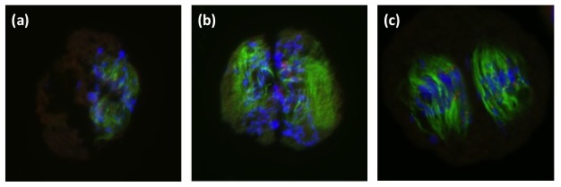
Figure 4
Immunostaining of the PMC in the 1H monosomic addition line. Cells were stained by anti-α-tublin antibody to detect spindles (green) and anti-CENH3 antibody to detect active centromeres (red). Chromsomes are counter-stained by DAPI. In panel (a) stretched chromosomes in the transition stage from MI to AI are visible but the spindle is so amorph as not to form bipolar spindle. Precocious cytokinesis is visible in (b) that is indicated by a ditch lacking spindle signals. In the irregular dyads, bipolar spindles seems to be organized in each cell (c).